
08 Feb MRI brain scan – elevated prolactin
The wonders of modern medicine, hey? I have been stressed out for most of the week, because I was scheduled to have an MRI scan of my brain this afternoon. The reason that I needed to get this done is because my prolactin is very high, and although a medication that I take elevates prolactin, my doctor wanted to rule out a tumour on my pituitary gland that can also cause elevated prolactin.
I was extremely dubious about the whole thing. It seems like it is highly unnecessary, when my prolactin will likely reduce as I reduce my medication further. But in my doctor’s own words – ‘it will ensure you don’t get sick, and that we don’t end up in court’. Whatever. As if I would sue.
Childhood phobia and a drip
In order to take an MRI scan of my pituitary gland, I needed to be injected with a contrast solution. When my doctor told me what the procedure would be, I was even more dubious about getting this scan done.
For a contrast MRI, first they insert a drip into your vein, and then you go into the MRI machine for about 15 minutes, while it does all sorts of different scans.
I hate blood tests at the best of times, but since a recent visit in hospital, where they were taking blood all the time, it is no longer a real phobia any more. It is still not something that I look forward to.
The radiographer came out while the nurse was inserting the drip and started talking to me, and asking me what was wrong. It was incredibly stressful. I just wanted to close my eyes and have it over with.
Another good tip, if you are going to get blood taken is to make sure that you drink lots of water, because that makes the veins stand out more.
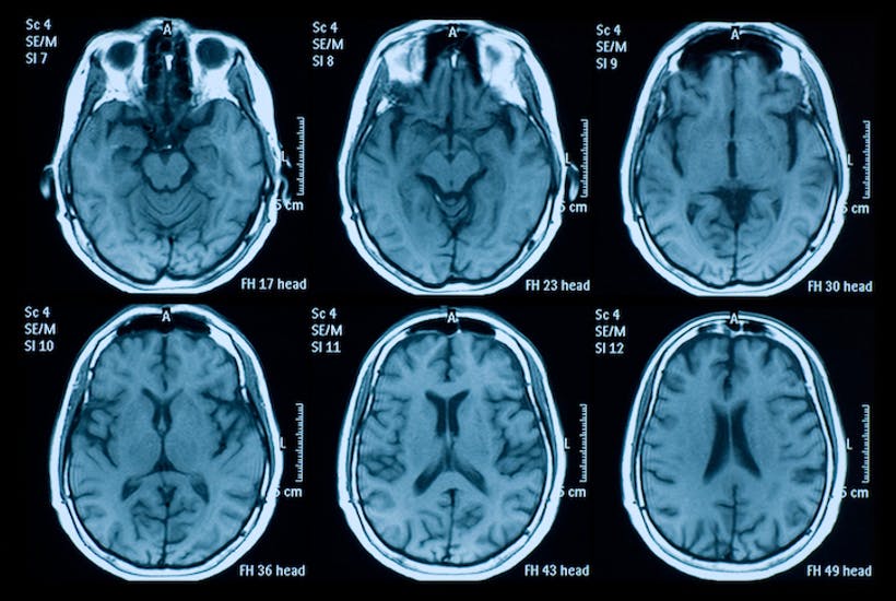
Magnetic Resonance Imagery (MRI) for elevated prolactin
I sat there with the drip in for about fifteen minutes and waited for the MRI machine to be free. Then they finally took me in. I was terrified. My doctor told me that people can get quite anxious and claustrophobic inside the machine. However it was not too bad.
They locked this kind of cage over my head, gave me earplugs, put padding around my head, gave me an alarm in my hand to press if I needed to come out for some reason (which was comforting), and wheeled me in.
It was actually not that stressful, and they played music – Florence and the Machine, so it was quite relaxing really. Also, there was a mirror in the top of the head cage, so that I could see the radiographer taking images, and he would talk to me through a microphone, periodically. So that I knew what was happening.
Contrast die risks
There are some risks with the contrast die that they inject into you when you have a contrast MRI. Apparently 1 in 365,000 go into anaphylactic shock, and it can cause, redness, itching, dizziness and nausea. None of these things happened to me. Phew!
I feel a bit light headed now, but I think I am fine to go out with my friends tonight. It was a pretty amazing experience, really. To think that they can see inside your brain like that. The machine is obviously pretty sophisticated.
And when the results come back I can rule out a brain tumour! Which is ideal 🙂 It is not GREAT about my elevated prolactin, because it can affect my heart and bones, but I think it will come down with my reduction in medication.
The best thing about the whole process is that I did it! After nightmares about it, and hoping they would ring and cancel and not wanting to talk about it all day. . . I did it! Hooray.
Samuel Pegram
Posted at 03:02h, 19 MarchMagnetic resonance imaging scanner does not use any radiation x-rays. To get pictures of your brain, the MRI scanner uses very strong magnet. It gives reliable scanning results of the brain and the spinal cord which is totally a safe procedure. However, it is an expensive procedure but these days’ medical experts are relying more on MRI scans.
https://www.rdch.org/
Pingback:vital sustenance Neurology appointment at St Vincent's Hospital - vital sustenance
Posted at 13:56h, 18 July[…] of medicine and to learn about what happens in a particular speciality. You might recall that I went in for a brain MRI earlier on in the year. The results came back, saying that ‘increased signal intensity is seen in the subcortical […]
Samuel Pegram
Posted at 01:32h, 17 DecemberMagnetic resonance imaging scanner does not use any radiation x-rays. To get pictures of your brain, the MRI scanner uses very strong magnet. It gives reliable scanning results of the brain and the spinal cord which is totally a safe procedure. However, it is an expensive procedure but these days’ medical experts are relying more on MRI scans.